
Over the past few years there has been quite the debate amongst many in the scientific community as to the extent of which the spine has the ability to adapt. While it is commonly accepted that the vertebral bones have the ability to adapt and become stronger/more dense with correctly dosed loading (a reason why powerlifters/weightlifters commonly have the highest bone mineral density in the world) there are still many questions to be answered when it comes to the intervertebral discs (IVDs).12-13
Recently some are claiming the IVDs can adapt to stress under load and become stronger and more resilient over time based on the findings from a recent study “Imaging of exercise-induced spinal remodeling in elite rowers” by Frenken et al 2022 in the Journal of Science and Medicine in Sport.1
However, is this study really showing the disc adapts?
Quick Synopsis
The paper supports:
- Cyclic loading enhances nutrient transport across the vertebral endplates to the nucleus
The paper does NOT support
- That the whole IVD responds to load similarly and therefore adapts in a beneficial manner.

In-Depth Synopsis
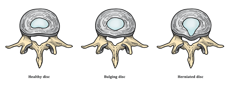
To understand the Frenken paper, you first need to comprehensively understand intervertebral disc anatomy and physiology.
The intervertebral disc has three components. They each have separate structure and function.
Intervertebral discs (IVDs) consist of
- an outer collagenous structure – the annulus fibrosus (AF),
- an inner nucleus pulposus (NP) and
- cartilage end plates (CEP) on the superior and inferior aspect.
Nucleus Pulposus
The NP has no blood or nerve supply. A normal NP is a gell-like structure that has a very high viscosity. It is composed of small proteins called proteoglycans and an intermolecular water gel held loosely by an irregular network of fine Type 2 collagen and elastin fibers. The major proteoglycan of the disc is aggrecan. Aggrecan, because of its high anionic glycosaminoglycan content (i.e., chondroitin sulfate and keratan sulfate), provides the osmotic properties needed to resist compression forces.2,3
The NP acts hydrostatically by transmitting pressure evenly to the annulus fibrosus and end plates in every direction according to Pascal’s principle.

On MRI, the hyperintense signal of the nucleus on T2- weighted images has been shown to correlate directly with the proteoglycan concentration in the NP. CEST imaging, as used in the Frenken study, is a non-invasive imaging technique that allows the determination of GAG content in IVDs.
Annulus Fibrosus (AF)
The annulus fibrosus is mostly avascular (very little blood supply) and only the outer third of the disc is innervated by nerves. It consists primarily of Type 1 Collagen fibers (remember the NP is Type 2). The rings of the annulus are called the lamellae. It is very different in structure, composition, and function to the NP. The very outer AF has a very limited blood supply, such that the annulus as an entire structure is considered to be avascular.
A tear in the AF does not adapt, and are not remodeled as in bone.8 Structural changes are irreversible because adult discs have limited healing potential.10
If you want to take a really deep dive into the science of the AF, check out this research article: “Annulus fibrosis cell phenotypes in homeostasis and injury: implications for regenerative strategies.”
Collagen turnover time in articular cartilage is approximately 100 years, and it may be even longer in the annulus.9,10 Proteoglycan turnover is possibly 20 years. Injuries that affect the inner annulus or endplate decompress the nucleus, and the slow “healing” processes are then overtaken by severe degenerative changes.7
Gross injuries to a disc never fully heal, never return to pre-injury status. Injury to the outer annulus fills with granulation tissue, only the outer few millimeters being bridged by scar tissue.6,7
While there is GAG content in the AF, there has been shown to be significantly lower GAG values in this part of the disc compared with the NP.4

Cartilaginous End Plates (CEP)
CEP is a layer of cartilage that lies in between the disc and the vertebral bone (think of it like the leather stretched over the top of a drum). It plays a crucial role in the maintenance of the mechanical environment as well as the proper nutrition of the avascular discs.
Frenken Paper
With our anatomy lesson out of the way, let’s now return to our topic for today. The Frenken paper investigates the Glycosaminoglycan (GAG) content of the IVD amongst elite rowers over the course of a training period. They found a temporary increase in this protein (via higher gagCEST values) and believed that to mean it is an indication of lumbar disc “remodeling effects” in response to training.

An interesting aspect of their findings was that the NP had almost a 2-FOLD increase in GAG compared to the AF. This increase however returned to normal levels after the rowing programming ceased. So while there was an increase in GAG noted in both areas of the IVD, there was significantly more increase in the NP (remember this as we’ll come back to this later).
So how does an increase in GAG content mean the IVD adapts?
Well, the results of this study show that repetitive loading of the spine supports nutrient transport across the vertebral endplates and an increase in GAG. Also, REDUCED GAG content is associated with IVD degeneration.(14).
So, if reduced GAG content is associated with disc degeneration, many believe the opposite can be true…an increase means the disc is positively adapting!
Not so fast…
This shows an association, not causation. The proof of nutrient availability isn’t the same as mechanism utility (simply put, it doesn’t mean the uptake of such nutrients is occurring).
Think of this like eating. You can eat a steak and have more protein circulating within the body, but that doesn’t mean you will automatically build muscle. GAG is only a protein within the extracellular matrix of the IVD that has been found to temporarily increase in response to loaded exercise and decrease (more so in the NP than the AF) in response to injury.5
While gagCEST of lumbar IVDs may be helpful to investigate early disc degeneration specifically looking at the NP, it is not a helpful way to determine physical changes in the AF.
It confirms the biological, and physiological, nature of cyclic compression for nuclear health.
If in fact the disc was truly adapting we would NOT see an overwhelming amount of research showing an extremely high prevalence of disc degeneration in rowers. (Sekine et al., 2014; Tomislav Smoljanovic et al., 2009; Hosea & Hannafin, 2012; A L Boland et al, 1991). In fact, the authors themselves said, “intensive athletic exercise at a professional level is associated with early degenerative changes, including disc herniation.”
With this understanding, we can NOT conclude whatsoever that a temporary increase in GAG content is proof that the IVD adapts positively to load. GAG content does not prevent disc degeneration.
We can established that yes the Frenken paper and many more (see below) do prove that the NP and the AF increase in GAG in response to certain stimuli. Loaded exercise, from rowing to walking, causes nutrient transfer across the vertebral endplates into the NP and AF (in fact, twice as much into the NP).
However, this does NOT prove adaptation. GAG is associated with degeneration, but not a direct causal relationship. Proving that nutrients are available and the mechanism and process involved in the uptake and application of those nutrients are two very different things. We have plenty of animal studies showing the AF does not heal effectively, and even when it may it’s with very poor quality. Scar tissue is laid down by the body that never really returns to full function and always remains at risk of future injury. Other studies show the inner portion of the annulus does not repair at all, and remains damaged long term.
The authors of the Frenken study conclude that the AF is half as “adaptive” in their study based on the findings that the NP had values of nearly double the AF. However, in context of the other studies discussed today, this is not valid. It is much more likely to adhere to maladaptation as confirmed by a TON of studies that specifically show rowers do not adapt to their training and have major degeneration in the spine and AF!
Studies showing the NP and AF adapt:
- DENG Min1, YUAN Jing2, CHEN Wei Tian1, Queenie CHAN3, James F GRIFFITH1, and WANG Yi Xiang1,#. Evaluation of Glycosaminoglycan in the Lumbar Disc Using Chemical Exchange Saturation Transfer MR at 3.0 Tesla: Reproducibility and Correlation with Disc Degeneration* (Can’t find this study anywhere officially) https://sci-hub.se/10.3967/bes2016.005
- Schleich, C., Müller-Lutz, A., Eichner, M., Schmitt, B., Matuschke, F., Bittersohl, B., Zilkens, C., Wittsack, H.-J., Antoch, G., & Miese, F. (2016). Glycosaminoglycan Chemical Exchange Saturation Transfer of Lumbar Intervertebral Discs in Healthy Volunteers. SPINE, 41(2), 146–152. https://doi.org/10.1097/brs.0000000000001144
https://sci-hub.se/10.1097/BRS.0000000000001144 - Schleich, C., Müller-Lutz, A., Matuschke, F., Sewerin, P., Sengewein, R., Schmitt, B., Ostendorf, B., Wittsack, H.-J., Stanke, K., Antoch, G., & Miese, F. (2015). Glycosaminoglycan chemical exchange saturation transfer of lumbar intervertebral discs in patients with spondyloarthritis. Journal of Magnetic Resonance Imaging, 42(4), 1057–1063. https://doi.org/10.1002/jmri.24877
https://sci-hub.se/10.1002/jmri.24877 - Belavý, D. L., Quittner, M. J., Ridgers, N., Ling, Y., Connell, D., & Rantalainen, T. (2017). Running exercise strengthens the intervertebral disc. Scientific Reports, 7(1). https://doi.org/10.1038/srep45975
So what do we know?
– The NP has significantly more GAG than the AF (Haneder et al., 2012)
– The AF is prone to maladaptive changes over time (Melrose et al., 1992)
– Discs don’t remodel like bone or heal like muscle/tendon tissues (Belavy et al, 2017)
– The AF has limited healing potential (Verzij| et al., 2000)
IN CONCLUSION
1.WE KNOW POOR TECHNIQUE UNDER LOAD CHALLENGES TISSUE TOLERANCE
2. WHEN TISSUE TOLERANCE IS EXCEEDED INJURY CAN OCCUR
3. CERTAIN POSITIONS ARE PARTICULARLY PROVOCATIVE TO THE DISC
4.PARTS OF THE DISC DO NOT HEAL OR ADAPT VERY WELL
5. STRATEGIES TO AVOID PROVOCATIVE MOVEMENT UNDER LOAD SHOULD BE EXPLORED

Until next time,

Dr. Aaron Horschig, PT, DPT, CSCS, USAW

Dr. Brogan Williams, PhD

Dr. Andrew Lock
References
- Frenken M, Schleich C, Radke KL, Müller-Lutz A, Benedikter C, Franz A, Antoch G, Bittersohl B, Abrar DB, Nebelung S. Imaging of exercise-induced spinal remodeling in elite rowers. J Sci Med Sport. 2022 Jan;25(1):75-80.
- Miles, D.E., Mitchell, E.A., Kapur, N., Beales, P.A. and Wilcox, RK. (2016) Peptide: Glycosaminoglycan Hybrid Hydrogels as an Injectable Intervention for Spinal Disc Degeneration. Journal of Materials Chemistry B, 4, 3225-3231.
- An, H.S., Anderson, P.A., Haughton, V.M., Iatridis, J.C., Kang, J.D., Lotz, J.C., et al. (2004) Introduction: Disc Degeneration: Summary. Spine, 29, 2677-2678.
- Haneder, S., Apprich, S.R., Schmitt, B., Michaely, H.J., Schoenberg, S.O., Friedrich, K.M., et al. (2013) Assessment of Glycosaminoglycan Content in Intervertebral S.
- Heüveldop et al. DOI: 10.4236/ijcm.2019.104020 269 International Journal of Clinical Medicine Discs Using Chemical Exchange Saturation Transfer at 3.0 Tesla: Preliminary Results in Patients with Low-Back Pain. European Radiology, 23, 861-868.
- Latz D, Frenken M, Schiffner E, Knautz M, Quante WA, Windolf J, Grassmann JP, Jungbluth P, Schleich C. Assessment of glycosaminoglycan content in intervertebral discs of patients with leg length discrepancy: A pilot study. J Orthop. 2019 Apr 8;16(5):363-367. doi: 10.1016/j.jor.2019.03.014. PMID: 31011249; PMCID: PMC6463742.
- Melrose J, Ghosh P, Taylor TK, et al. A longitudinal study of the matrix changes induced in the intervertebral disc by surgical damage to the annulus fibrosus. J Orthop Res 1992;10:665–76.
- Kaigle AM, Holm SH, Hansson TH. 1997 Volvo Award winner in biomechanical studies. Kinematic behavior of the porcine lumbar spine: A chronic lesion model. Spine 1997;22:2796–806.
- Roughley PJ. Biology of intervertebral disc aging and degeneration: Involvement of the extracellular matrix. Spine 2004;29:2691–9
- Verzijl N, DeGroot J, Thorpe SR, et al. Effect of collagen turnover on the accumulation of advanced glycation end products. J Biol Chem 2000;275: 39027–31.
- Adams MA, Freeman BJ, Morrison HP, et al. Mechanical initiation of intervertebral disc degeneration. Spine 2000;25:1625–36
- Dickerman RD, Pertusi R, Smith GH. The upper range of lumbar spine bone mineral density? An examination of the current world record holder in the squat lift. Int J Sports Med. 2000 Oct;21(7):469-70.
- Walters PH, Jezequel JJ, Grove MB. Case study: Bone mineral density of two elite senior female powerlifters. J Strength Cond Res. 2012 Mar;26(3):867-72.
- Silagi, E. S., Shapiro, I. M., & Risbud, M. V. (2018). Glycosaminoglycan synthesis in the nucleus pulposus: Dysregulation and the pathogenesis of disc degeneration. Matrix Biology: Journal of the International Society for Matrix Biology, 71-72, 368–379. https://doi.org/10.1016/j.matbio.2018.02.025
Other interesting studies to read:
–Under Pressure: Mechanical Stress Management in the Nucleus
–Stress for Vertebral Bodies and Intervertebral Discs with Respect to Squatting Depth
The article mentions that cyclic loading enhances nutrient transport across the vertebral endplates to the nucleus. Does this increased nutrient transport directly correlate with improved disc health and resilience, or are there other factors at play that could negate the benefits?
Instead of racing to the finish line, the Drift Hunters goal is to maintain smooth drifts through corners while earning points based on angle, speed, and drift duration.
I recently tried https://faceshapedetector.ai/ and found it really simple and quick to use—it gives a nice breakdown of your face shape in just a few seconds!
Really insightful breakdown of a topic that’s often oversimplified in the fitness space. I especially appreciated how you clarified that while cyclic loading may improve nutrient transport, it doesn’t necessarily mean the intervertebral disc adapts in a stronger or more resilient way
This analysis clearly distinguishes correlation from causation, emphasizing that increased nutrient presence does not guarantee adaptation, and highlights evidence showing spinal loading may lead to degeneration rather than meaningful tissue recovery.
crossy road
Very interesting breakdown 🧠 It shows how discs respond to load, but doesn’t fully prove true adaptation. Great reminder that technique and load management matter most 💪 http://doraemonsx.com/
This is fascinating! If intervertebral discs do adapt to load, what implications does this have for strength training? Could it change how we approach injury prevention in athletes?
Could it change how we approach injury prevention in athletes cookie clicker free?
The analogy about eating a steak perfectly captures the flaw in the adaptation argument—just because the building blocks (GAGs) are present doesn’t mean the body is actually building a more resilient “wall” in the annulus fibrosus. The fact that these levels return to baseline after training stops really suggests it’s more of a transient physiological fluctuation than a permanent structural remodeling like we see in bone mineral density. I love video compressor
The way intervertebral discs respond to mechanical stress is closely tied to hydration, daily activity, and gradual load distribution. Over time, consistent movement patterns can influence how spinal structures maintain flexibility and resilience under pressure. Additional structured information on related structural and data-based records can be reviewed through Volusia Property Insights for broader reference. Maintaining awareness of how systems adapt under repeated load helps in understanding both biological and analytical perspectives.
Research on intervertebral discs suggests they can respond to mechanical loading over time, although the extent of adaptation may depend on factors such as age, load intensity, and recovery periods. Understanding structural changes often requires access to detailed records and documentation, which is why resources like Hawaii records lookup can be valuable for organized information research. Maintaining balanced movement, proper posture, and gradual load progression may also support long-term spinal health. It would be interesting to see more studies examining how different loading patterns influence disc resilience.
You really need to try this https://bethall-au.com/ , for me this a perfect experience to spin your time 24/7. Just join and share your experience